
Managing acute abdominal pain in a pregnant patient represents one of the most intricate and high-stakes challenges in modern emergency medicine, requiring a delicate balance between rapid diagnostic accuracy and the preservation of fetal development. Medical professionals often find themselves at a crossroads where the necessity of immediate intervention must be weighed against the potential dangers of ionizing radiation exposure. This diagnostic environment is complicated by the fact that the health of two separate but deeply connected patients must be prioritized simultaneously, often under significant time pressure and clinical uncertainty. To mitigate these risks, modern clinical pathways are evolving to integrate more sophisticated and non-invasive imaging technologies that provide the clarity needed for life-saving decisions. The shift toward evidence-based imaging strategies highlights a broader commitment to maternal-fetal safety, ensuring that physicians can act with confidence rather than caution when faced with ambiguous clinical presentations in the emergency department.
The Diagnostic DilemmNavigating Obstacles in Maternal Health
Physiological adaptations occurring during pregnancy frequently obscure the clinical picture, making it difficult for even experienced physicians to distinguish between normal discomfort and a surgical emergency. The expansion of the uterus naturally displaces internal organs, which can lead to atypical pain patterns and render standard physical examinations less reliable than in the non-pregnant population. For instance, the upward migration of the appendix as the pregnancy progresses can cause pain to manifest in the upper right quadrant rather than the lower right, potentially misleading the primary care team. Furthermore, laboratory findings that would typically indicate a severe infection, such as an elevated white blood cell count or an increased heart rate, are often within the normal physiological range for an expectant mother. These overlaps create a complex diagnostic landscape where clinical signs alone are insufficient to confirm a specific diagnosis or rule out a life-threatening complication.
The uncertainty surrounding these physical manifestations often culminates in a phenomenon known as diagnostic hesitancy, where medical providers delay necessary testing due to excessive concerns over fetal safety. While this caution is rooted in the principle of doing no harm, an overly conservative approach can lead to catastrophic results if a serious condition remains undetected for an extended period. Untreated abdominal infections or infarctions significantly heighten the probability of systemic maternal illness, sepsis, and spontaneous fetal loss. Because the risks of a missed diagnosis are often far greater than the risks of modern imaging, there is an urgent need for standardized protocols that emphasize early and effective evaluation. Addressing these delays requires a systemic shift in how hospitals manage acute pain, moving toward rapid access to advanced diagnostics that can definitively identify the source of distress without subjecting the developing fetus to unnecessary biological stress or long-term risks.
Traditional Modalities: Evaluating the Efficacy of Conventional Tools
Ultrasonography serves as the initial line of defense in the evaluation of the maternal abdomen because of its accessibility, portability, and complete lack of ionizing radiation. It remains the preferred screening tool for identifying common issues such as gallstones, kidney obstructions, or pelvic masses that might be contributing to a patient’s acute discomfort. However, the utility of ultrasound is frequently limited by the advanced gestational age of the fetus or the specific body habitus of the patient, which can create significant acoustic shadows. As the second and third trimesters progress, the sheer size of the gravid uterus can physically block the visualization of deeper retroperitoneal structures and the intestinal tract. Consequently, radiologists often encounter inconclusive results, where the target organ, such as the appendix, simply cannot be visualized. This limitation necessitates a more robust secondary imaging option to provide the diagnostic certainty required for surgical planning.
Computed Tomography has long been considered the gold standard for diagnosing acute abdominal issues in the general population, yet its application during pregnancy remains restricted due to radiation concerns. The ionizing radiation inherent in CT scans poses theoretical risks to the fetus, including potential developmental abnormalities or an increased risk of malignancies. While the medical community has developed low-dose CT protocols specifically for use in pregnant women, these are generally reserved for extreme trauma or life-threatening emergencies where maternal survival is at immediate risk. Even with these precautions, the use of intravenous iodinated contrast agents adds another layer of complexity, as these substances can cross the placenta and potentially affect fetal thyroid function. Therefore, while CT remains a powerful and fast diagnostic tool, it is increasingly viewed as a last resort in the prenatal context, used only when other modalities have failed to clarify a critical clinical situation.
Magnetic Resonance: Utilizing Advanced Protocols for Fetal Safety
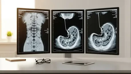
Magnetic Resonance Imaging has redefined the standard of care for pregnant patients by providing a high-resolution window into the abdomen without the hazards associated with ionizing radiation. By utilizing strong magnetic fields and radiofrequency pulses, MRI generates incredibly detailed cross-sectional images that surpass the clarity of ultrasound while maintaining a superior safety profile compared to CT scans. Current medical guidelines frequently recommend the use of 1.5-Tesla scanners for these patients, as this field strength offers an ideal balance between high image quality and established safety data. This technology is particularly valuable when the initial ultrasound is inconclusive, as it allows radiologists to see through the obstructions caused by the growing uterus. The ability to perform multiplanar imaging enables the medical team to visualize the appendix and gallbladder with exceptional precision, facilitating a definitive diagnosis that can guide subsequent medical or surgical interventions.
Recent advancements in specialized MRI sequences, such as Diffusion-Weighted Imaging, have further expanded the diagnostic capabilities of this modality without requiring external contrast agents. By measuring the random motion of water molecules within the body, these sequences can pinpoint areas of restricted movement that typically correspond with inflammation, abscess formation, or malignant growth. This functional information is critical because it allows radiologists to identify active disease processes at a cellular level, providing a layer of detail that traditional anatomical imaging might miss. Since many gadolinium-based contrast agents are avoided during pregnancy due to their ability to cross the placenta and remain in the amniotic fluid, these non-contrast techniques are essential. They provide the clinical team with necessary insights into the severity of an infection, thereby accelerating the time to treatment and reducing the overall duration of the hospital stay for the expectant mother.
Institutional Progress: Shaping Future Standards in Perinatal Care
The broader adoption of specialized imaging protocols signaled a significant turning point in the management of obstetric patients, ensuring that safety and accuracy were no longer mutually exclusive. Medical institutions that prioritized the installation of rapid-access MRI for their emergency departments observed a measurable decrease in unnecessary surgical interventions and improved overall maternal outcomes. Healthcare providers successfully navigated the complexities of prenatal diagnostics by moving away from outdated reliance on clinical observation alone toward a more proactive, technology-driven approach. This transition solidified the role of the radiologist as a critical consultant in the perinatal care team, providing the objective data needed to resolve clinical uncertainty. By standardizing these safety-first imaging practices, the medical community established a foundation for future innovations that will continue to protect the most vulnerable populations across the healthcare spectrum.
Hospitals that integrated these protocols into their core emergency workflows successfully bridged the gap between radiology and obstetrics, resulting in a more cohesive patient journey. This evolution in care suggested that the primary solution for reducing diagnostic ambiguity lay in the consistent availability of advanced MRI resources. Moving forward, the emphasis was placed on training specialized technicians and radiologists to recognize the unique markers of prenatal pathology in non-contrast sequences. These systematic changes proved that institutional commitment to sophisticated imaging could significantly lower the risks of maternal morbidity and unnecessary operative procedures. Ultimately, the transition to high-resolution, radiation-free diagnostics became the definitive strategy for ensuring safety and precision in maternal emergency care. The successful implementation of these specialized diagnostic pathways served as a benchmark for modernizing perinatal medicine and improving clinical standards.